Keratoconus
Understanding Keratoconus
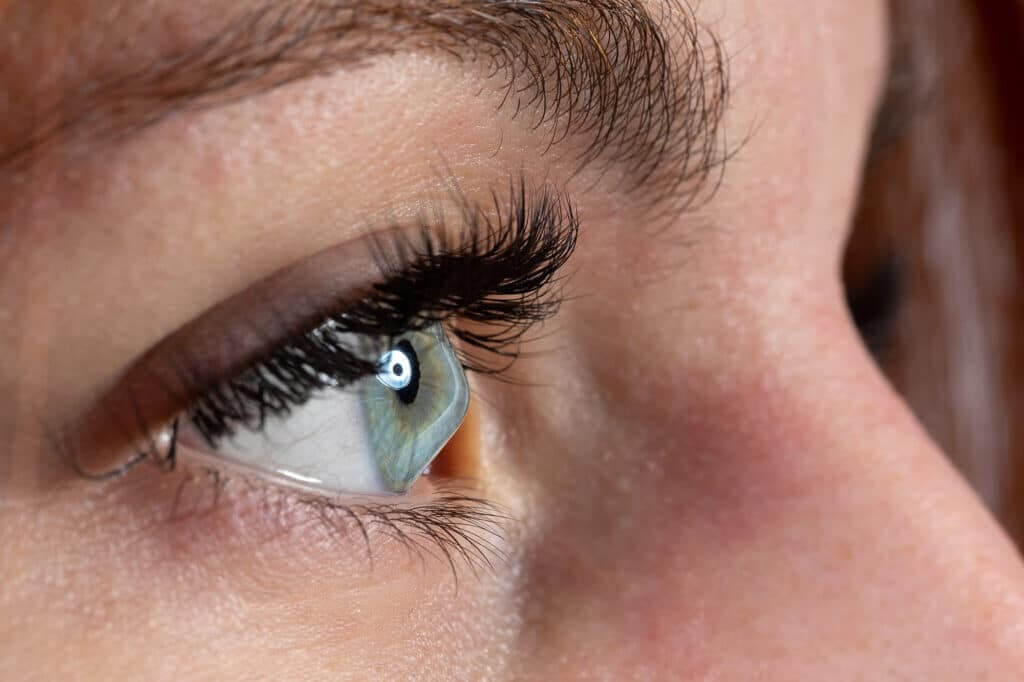
Keratoconus is a vision disorder of the eye in which the cornea (the clear front of the eye) becomes weaker and thinner, taking on a more cone-like shape than its normal dome-like curve. Patients with keratoconus gradually develop a progressively blurrier vision. Symptoms typically begin in the late teens or late twenties.
Because the cornea bulging has an irregular shape, glasses and conventional soft contact lenses cannot provide clear vision.
Causes of Keratoconus
The exact causes of keratoconus are uncertain, but various studies have shown contributions of genetic, environmental, and cellular factors.
The root cause is thought to be weakening of the fine structures that typically strengthen the cornea, which leads to bulging of the cornea in keratoconus.
Treatment for Keratoconus in Orange County, CA
In the early stages of keratoconus..
Glasses or soft contact lenses can correct mild astigmatism.
In moderate stages of keratoconus..
Rigid gas-permeable contact lenses (RGPs) provide a good level of vision correction but do not slow the progression of the condition. These lenses create a smooth front surface for the eye.
In advanced stages of keratoconus..
In the past, when rigid contact lenses failed, the only other option for treating keratoconus was a corneal transplant. Corneal transplant is often needed in very advanced keratoconus when other treatments have failed. This procedure requires a recovery period of 6-18 months before a stable vision can be achieved. After recovery, many patients still require rigid contact lenses to see well.
Corneal Cross-Linking (CXL)
Cross-linking is a non-invasive procedure designed to strengthen a weakened cornea, most commonly used to treat keratoconus. It works by applying special eye drops (riboflavin) to the cornea, followed by controlled UV light exposure. This process helps “cross-link” the collagen fibers in the cornea, making it more stable and less likely to bulge over time.
Benefits of Cross-Linking:
- Slows or stops progression of keratoconus
- Helps preserve your vision
- Safe, outpatient procedure

AVAGEN™ – THE GENETIC EYE TEST
AvaGen™ is the genetic eye test that provides answers on your risk for keratoconus and other corneal diseases—helping you and your doctor make confident eye care decisions now.
The AvaGen™ test is unique because it’s:
Personalized
AvaGen™ uses DNA to accurately assess your individual genetic risk or likelihood of keratoconus, and determine if you have a corneal dystrophy.
Preemptive
As a genetic test, AvaGen™ relies on your DNA and not physical changes in your eye, so you may be tested before symptoms of keratoconus or corneal dystrophies occur, allowing for earlier detection and proactive management and treatment.
Painless
A DNA sample is simply taken from the inside of your cheek with a cotton swab.
WHY IS AVAGEN™ IMPORTANT?
Genetic eye conditions including keratoconus and corneal dystrophies are progressive, meaning they will get worse over time if left untreated. In the past, keratoconus and corneal dystrophy eye diseases could only be diagnosed by a doctor physically examining the shape and appearance of your cornea, meaning conditions often went undiagnosed, worsened over time, and permanently affected vision.
Now, AvaGen™ can help your doctor take immediate actions to help protect and preserve your vision or to evaluate treatment decisions, including whether or not you can undergo vision-correcting procedures such as LASIK.
With AvaGen™, you will have the peace of mind of knowing you’re doing the utmost to protect your vision.
HOW IS THE AVAGEN™ TEST PERFORMED?
The AvaGen™ test only requires a simple, in-office cheek swab to gather a sample of your DNA. The test is then sent to a special lab for analysis. After processing, your doctor will inform you of the results, answer questions, and discuss potential next steps.
Comprehensive genetic counseling sessions are available for patients who have undergone testing with the AvaGen™ genetic eye test. Genetic counselors are available to help you understand your genetic eye test results and implications for your family members.
WHY WOULD MY DOCTOR RECOMMEND AVAGEN™?
- You have a family history of certain corneal problems or corneal dystrophy, vision changes, or a family member who had a corneal transplant for unknown reasons.
- Your doctor sees corneal thinning, or suspicious signs such as protein deposits on your corneas.
- You have high or progressive nearsightedness or astigmatism.
- You are considering refractive surgery such as LASIK.