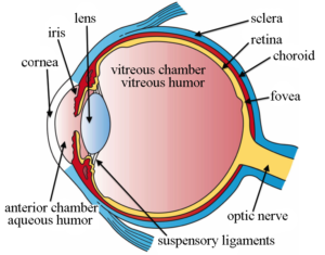
 Even though the eye is small, only about 1 inch in diameter, it serves a very important function – the sense of sight. The eye is one of the most complex organs in the human body. It is made up of many distinct parts working in unison together and in order for the eye to work at its best, all parts must work well collectively.
Let’s look at a few of the main parts of this vital organ:
Anterior Chamber: The space between the cornea and the iris, filled with the aqueous humor.
Choroid: The vascular layer of the eye, containing connective tissue. Nutrition of the eye is dependent upon blood vessels in the choroid.
Ciliary Body: the part of the eye that connects the iris to the choroid.
Cornea: The clear, dome-shaped tissue covering the front of the eye.
Fovea: A tiny pit located in the macula of the retina that provides the clearest vision of all.
Iris: The colored part of the eye that controls the amount of light that enters the eye by changing the size of the pupil.
Lens: A crystalline structure located just behind the iris – it focuses light onto the retina.
Macula: The small area at the center of the retina responsible for what we see straight in front of us.
Optic Nerve: The nerve that transmits electrical impulses from the retina to the brain.
Pupil: The opening in the center of the iris. It changes size as the amount of light changes.
Retina: Light-sensitive tissue that lines the back of the eye. It contains millions of photoreceptors (rods and cones) that convert light rays into electrical impulses that are relayed to the brain via the optic nerve.
Vitreous Gel: A thick, transparent liquid that fills the center of the eye. It is mostly water and gives the eye its form and shape.
Our eyes are vital for seeing the world around us. Keep them healthy by maintaining regular vision exams. Contact Harvard Eye Associates at 949-951-2020 or harvardeye.com to schedule an appointment today.


